


Retinal detachment
Retinal detachment is an emergency: the retina separates from the wall of the eye and stops working.
Read article ↗
Eye conditions · 3 MIN READ
WHAT YOU NEED TO KNOW
The macula is the central part of the retina, responsible for the detailed vision we use to read, recognise faces and drive. When a film forms over the macula, it can contract and wrinkle the retina, like crumpled cellophane. Hence the names epiretinal membrane, macular pucker or cellophane maculopathy.
In most people it is related to ageing of the vitreous, the gel that fills the eye. With age, the vitreous separates from the retina and some cells may remain on the surface of the macula and form the membrane. It is more common after the age of 50. It can also follow a retinal detachment or tear, eye surgery, inflammation, injury or retinal vascular disease such as diabetic retinopathy.
THE MACULA IN CROSS-SECTION
How the membrane distorts vision
The membrane contracts and wrinkles the retina, like crumpled cellophane.
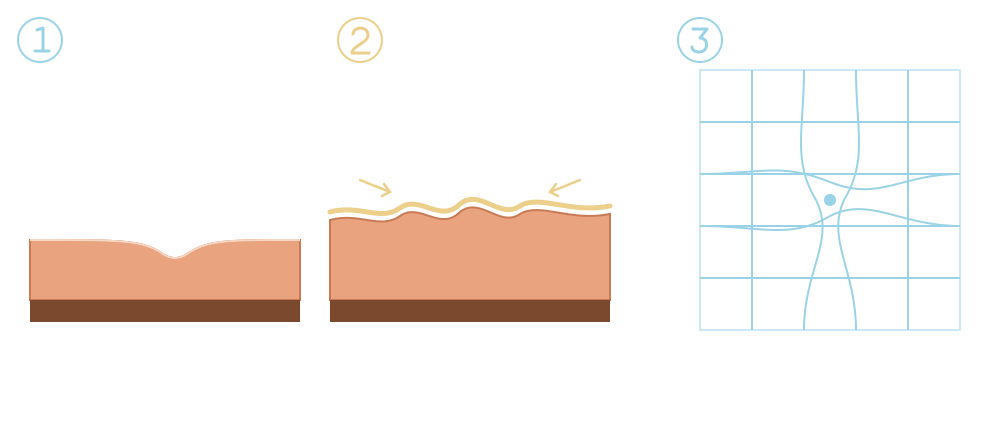
The macula has a small central dip, the fovea, where vision is sharpest.
The film on the surface pulls the retina, which becomes thick and folded.
Straight lines look wavy or bent, especially in the centre.
It usually progresses slowly. Many membranes stay stable for years and they rarely go away on their own.
The membrane can be seen when the back of the eye is examined. Optical coherence tomography (OCT) is the key test: it shows a cross-section of the membrane, the thickening and the folds of the retina, and allows changes to be followed and the timing of surgery to be decided. The Amsler grid helps you monitor distortion at home.
There are no eye drops or tablets that make the membrane go away. If symptoms are mild, the usual approach is to monitor it with appointments and OCT. New glasses can improve sharpness but do not correct distortion.
When vision or distortion interferes with daily life, surgery is proposed: vitrectomy with removal of the membrane. The surgeon removes the vitreous and, with very fine forceps, peels the membrane off the surface of the macula. Surgery is usually done under local anaesthesia.
If you suddenly notice many floaters, flashes of light or a shadow in your field of vision, seek urgent assessment: they may indicate a retinal tear or detachment.
This information does not replace an individual medical assessment. Follow the advice of the team looking after you.
Clinical responsibility: Dr. Nuno Lopes · Ophthalmology
Page update:
This date refers to the page presentation and organisation.
CONTINUE READING

Retinal detachment is an emergency: the retina separates from the wall of the eye and stops working.
Read article ↗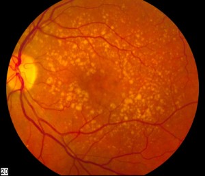
AMD affects the macula, the part of the retina responsible for central, detailed vision.
Read article ↗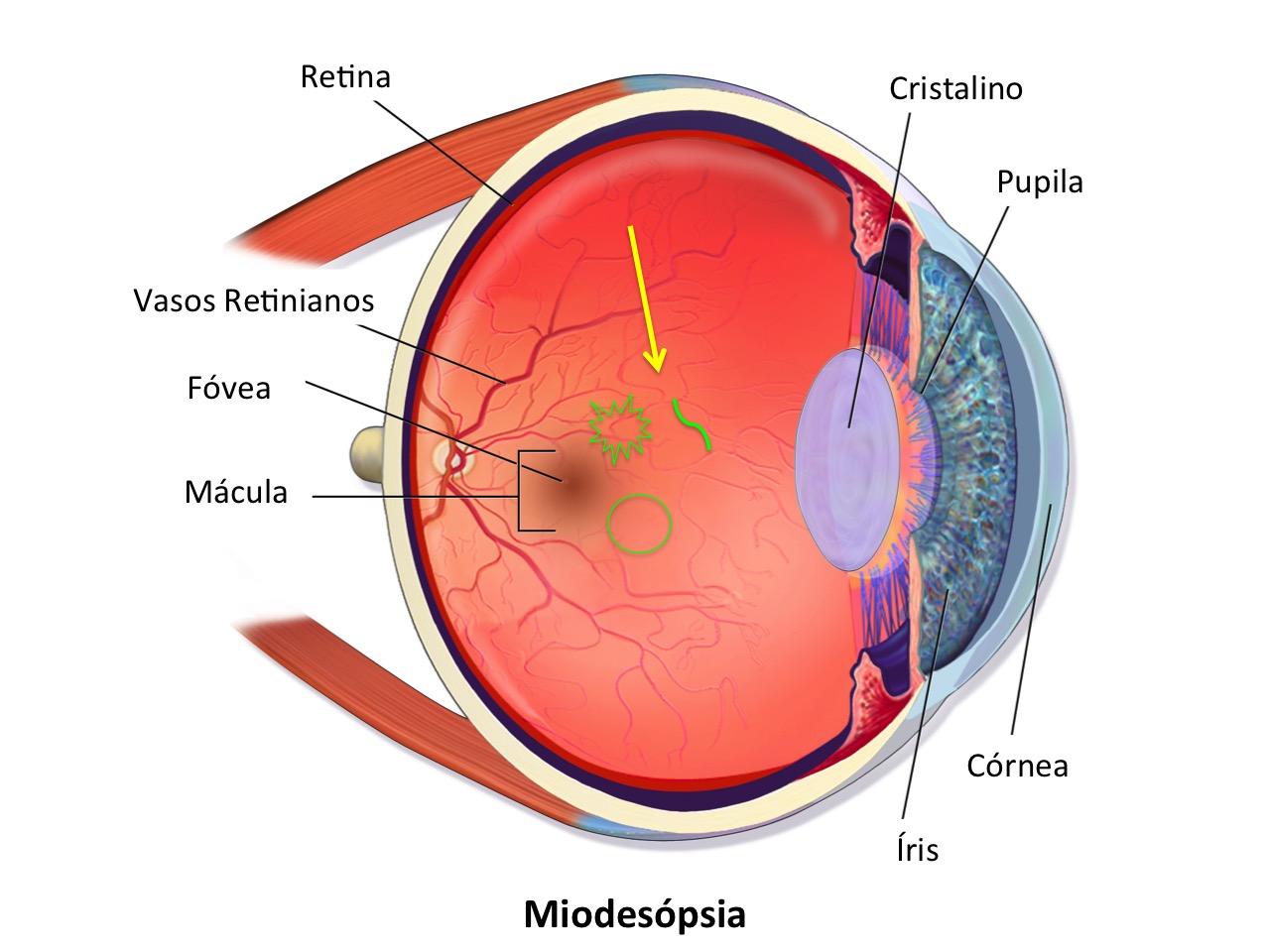
Floaters are shadows that move across the field of vision and are very common with age.
Read article ↗