


INFORMATION TO CARE FOR YOUR VISION
Questions and Guidance
Answers to your questions and practical advice, organised by topic.
25 artigos
Appointments and follow-up
 At what age and how often should I see an ophthalmologist?
Newborns are usually examined by their paediatrician.
Vision screening continues to be carried out regularly by the paediatrician between 6 months and one year of age.
If there is no suspected eye disease requiring earlier referral, the first examination by an ophthalmologist usually takes place between 2 and 3 years of age, followed by a preschool examination.
Healthy adolescents and adults generally have regular reviews every 2 years, changing to annual reviews from the age of 40.
When eye disease is present, the ophthalmologist determines the follow-up intervals according to the condition and its stage.
Some systemic diseases may require more frequent follow-up, for example:
• DIABETES
• CONNECTIVE TISSUE DISEASES (LUPUS; RHEUMATOID ARTHRITIS; SJÖGREN SYNDROME)
• THYROID DISEASES
A family history of eye diseases such as glaucoma should also prompt earlier monitoring.
This information does not replace an individual medical assessment. Follow the advice of the team looking after you.
Open article on its own pageGlaucoma
 Ocular hypertension and glaucoma: what is the difference?
High eye pressure is not the same as glaucoma. Understand the difference, the tests and the importance of personalised follow-up.
Read article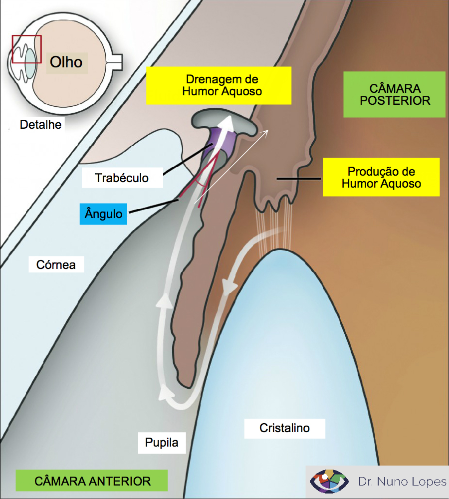 What is intraocular pressure or eye pressure?
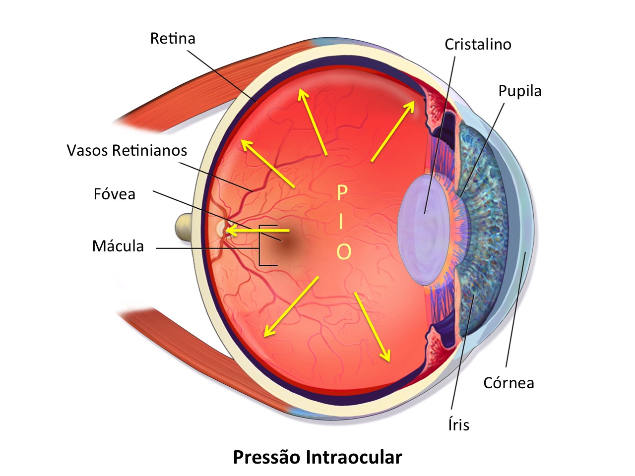
Intraocular pressure (IOP), or eye pressure, measures the pressure or force inside the eyeball.
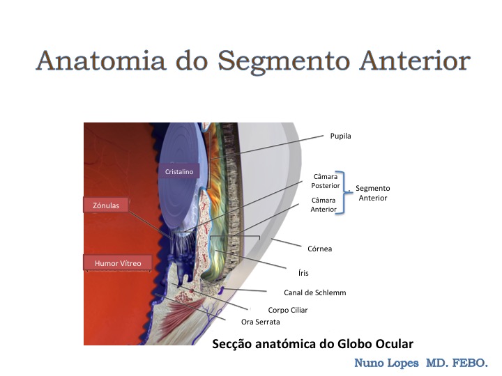
Its level reflects the balance between the production and drainage of aqueous humour, the fluid produced by the ciliary body at the junction between the front and back parts of the eye.
Normal values vary between populations and individuals. Values above 21 mmHg are generally considered ocular hypertension, but conclusions should not be drawn from an isolated IOP reading. The average value in the Caucasian population is 16 mmHg.
IOP fluctuates by up to about 5 mmHg during the day in healthy individuals and may fluctuate more in patients with glaucoma. Several factors influence IOP fluctuation, including seasonal changes and posture.
Higher IOP is associated with a greater risk of developing glaucoma and of its progression.
This information does not replace an individual medical assessment. Follow the advice of the team looking after you.
Open article on its own page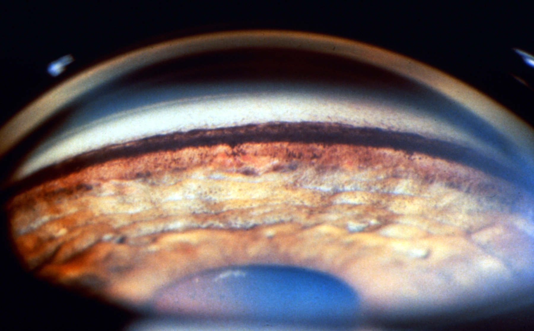 How does open-angle glaucoma differ from angle-closure glaucoma?
Glaucoma is a progressive optic neuropathy that produces characteristic anatomical changes in the optic nerve and corresponding loss of visual field sensitivity.
The main risk factor for its development is ocular hypertension, although normal-tension glaucoma also occurs.
Intraocular pressure reflects the balance between production and drainage of aqueous humour. As its production rate is reasonably stable, ocular hypertension usually results from insufficient drainage.
The main outflow route for aqueous humour is through the trabecular meshwork at the angle where the iris inserts into the corneoscleral region.
It is called open-angle glaucoma when access to the angle appears unobstructed, and narrow-angle or angle-closure glaucoma when obstacles prevent aqueous humour from adequately reaching the trabecular meshwork.
Open-angle glaucoma is much more common than narrow-angle glaucoma. The latter, however, is generally more destructive and, despite being less common, accounts for almost 50% of glaucoma blindness.
Angle assessment is essential when glaucoma is suspected or diagnosed.
The best method of assessment is usually gonioscopy.
Additional tests such as optical coherence tomography (OCT) and ultrasound biomicroscopy (UBM) are also useful for this assessment.
This information does not replace an individual medical assessment. Follow the advice of the team looking after you.
Open article on its own page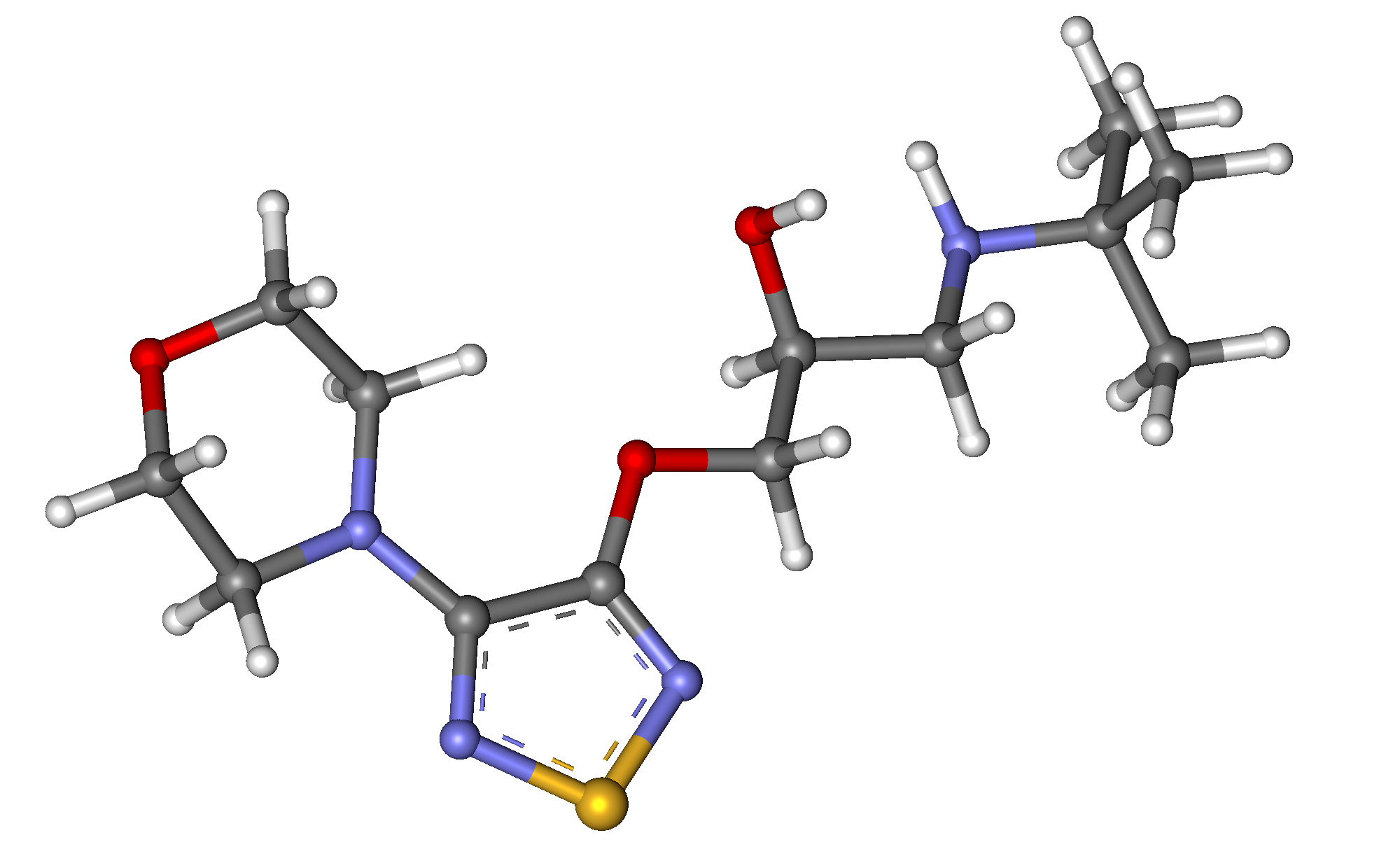 How is glaucoma treated?
As there is no definitive cure, the main aim of glaucoma treatment is to reduce risk factors for progression.
Of all known factors, raised intraocular pressure (IOP) is the most relevant, making pressure reduction the main target of treatment.
IOP can be lowered with topical medication in the form of eye drops and, less commonly, tablets. There are several classes of medication, some more appropriate for particular glaucoma subtypes.
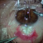
Eye drops are usually used as first-line treatment, although laser treatments are increasingly used. The most common include selective laser trabeculoplasty (SLT) to reduce IOP in open-angle glaucoma and iridotomy, typically used in narrow-angle glaucoma.
When necessary, surgery may also be performed to improve IOP control. The main reasons for considering laser treatment or surgery are:
1 – Insufficient IOP control with topical medication
2 – Significant disease progression despite maximum topical treatment
3 – Eye drop intolerance due to allergy or toxicity
4 – An expressed wish to reduce the need for pressure-lowering eye drops
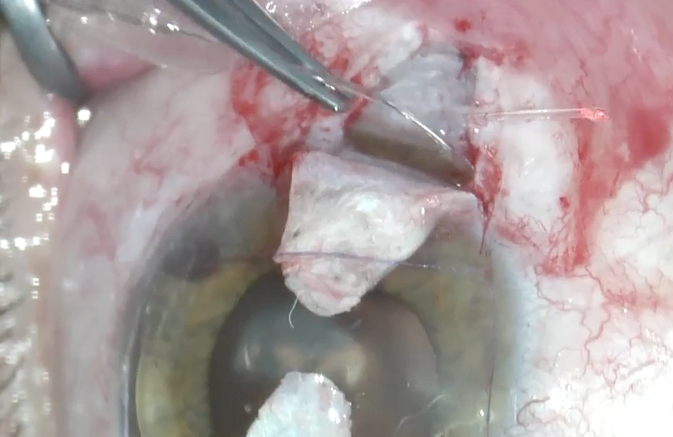
Several surgical alternatives are available today, and no single technique should be used across all types of glaucoma.
The best option usually depends on the type of glaucoma and the condition of the eye. Your ophthalmologist can advise which is most suitable for you.
Canaloplasty
Deep sclerectomy
Trabeculectomy
Lens surgery
Learn more:
 Watch video on YouTube ↗Original video in Portuguese
This information does not replace an individual medical assessment. Follow the advice of the team looking after you.
Open article on its own page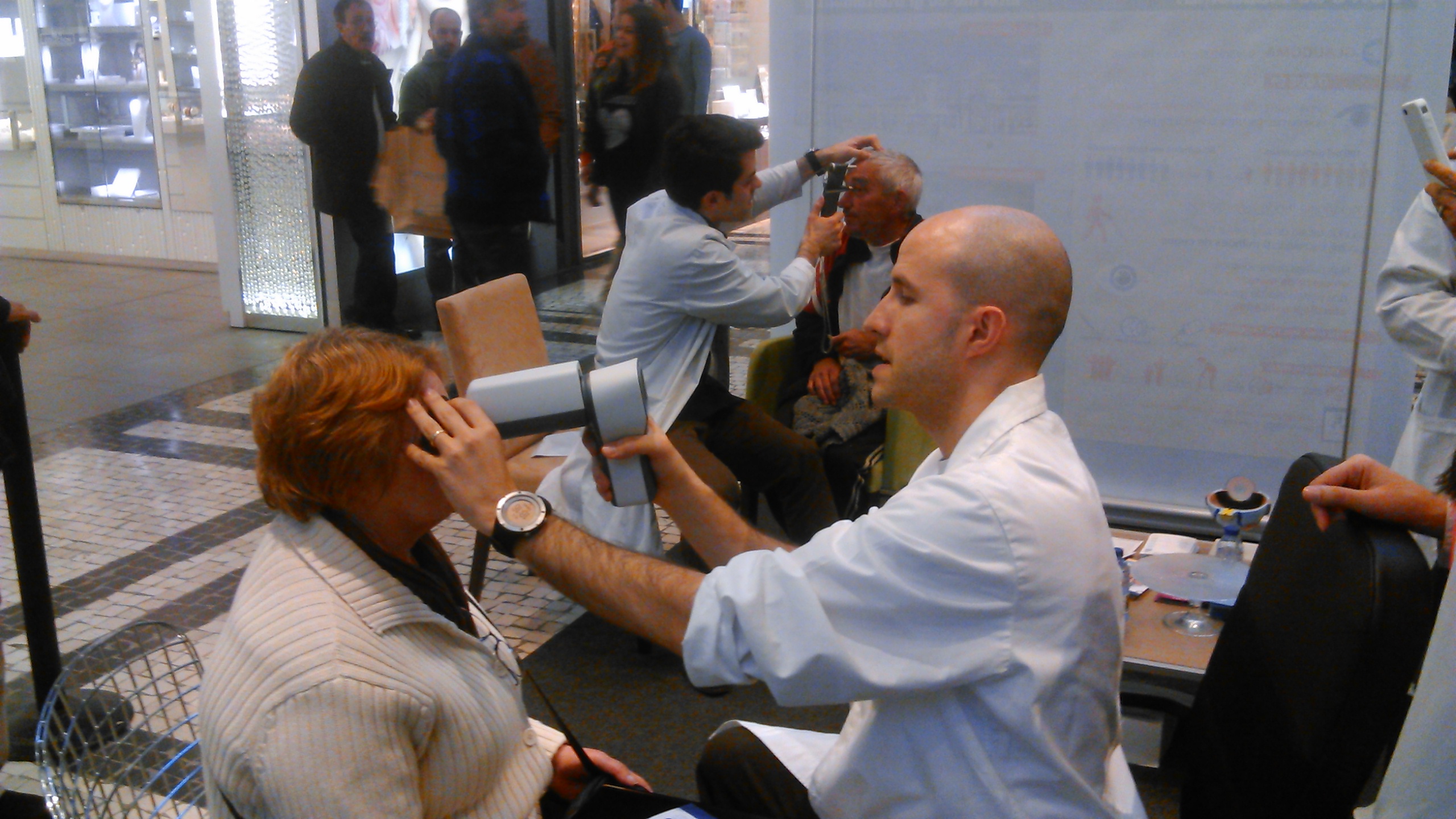 How is glaucoma detected?
The most common types of glaucoma, open-angle glaucoma, are usually symptom-free. Early diagnosis is therefore generally made by an ophthalmologist during screening or a routine examination.
Suspicion should be higher when one or more risk factors for the disease are present.
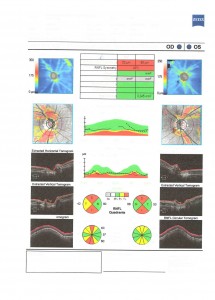
Very advanced glaucoma can be detected promptly with few additional tests. In early stages, however, optical coherence tomography (OCT), perimetry (visual fields), gonioscopy (angle assessment) and pachymetry are usually performed.

Computerised static perimetry
Optic disc OCT
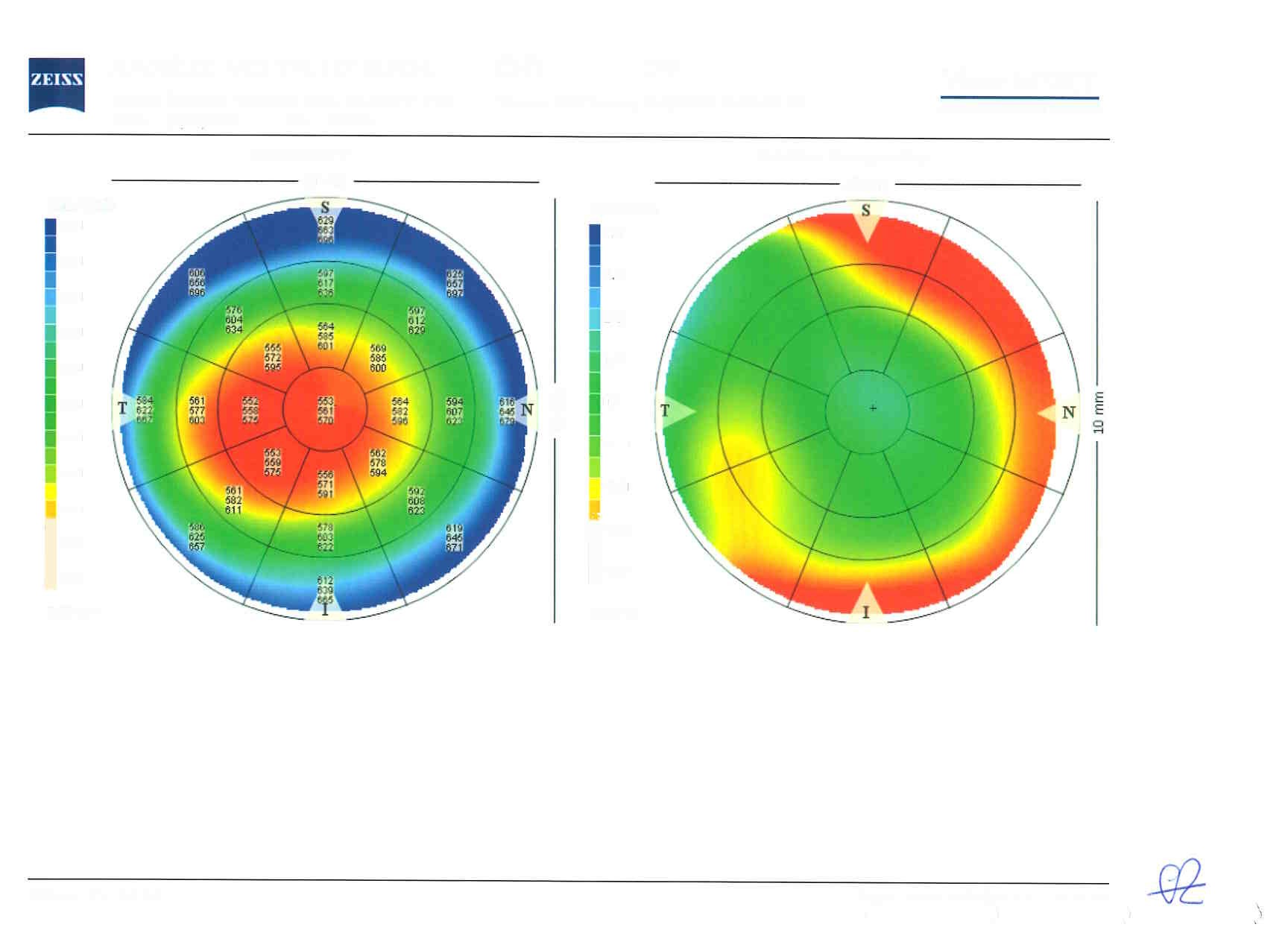
Pachymetry
Learn more:
Watch video on YouTube ↗Original video in PortugueseThis information does not replace an individual medical assessment. Follow the advice of the team looking after you.
Open article on its own pageWhat is glaucoma?
Glaucoma is a disease of the optic nerve.
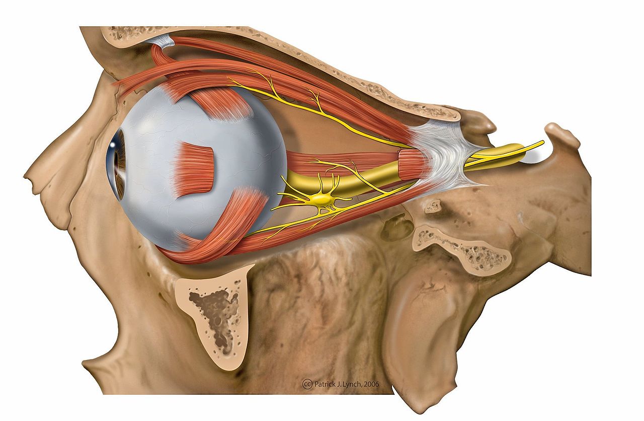
The posterior course of the optic nerve
The optic nerve (ON) carries images captured by the eye to the brain. As nervous tissue, it consists of millions of cells whose health is essential. Some natural cell loss is usual throughout life, but glaucoma causes excessively rapid loss that can threaten the patient's visual independence.

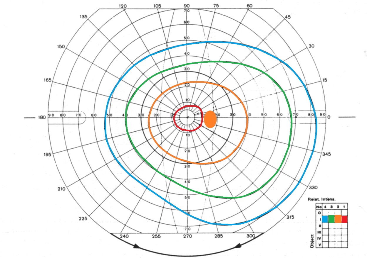
Illustration of visual field loss in advanced glaucoma
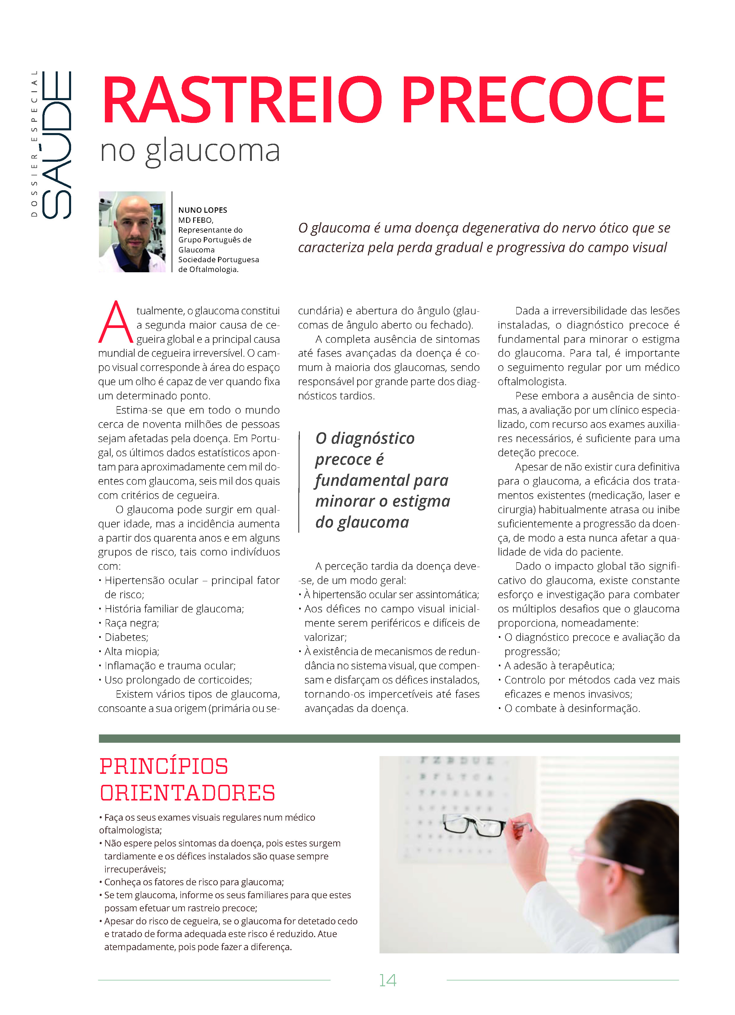
Glaucoma is a degenerative disease of the optic nerve characterised by gradual, progressive visual field loss. It is currently the second leading cause of blindness overall and the leading cause of irreversible blindness worldwide.
The visual field is the area of space an eye can see while looking steadily at a particular point.
An estimated ninety million people worldwide are affected by the disease.
In Portugal, the latest figures suggest approximately one hundred thousand people with glaucoma and six thousand who are blind because of it.
Glaucoma can occur at any age, but its incidence increases from the age of forty and in certain risk groups, including people with:
• Ocular hypertension — the main risk factor
• A family history of glaucoma
• Black or Asian ancestry
• Diabetes
• High myopia
• Eye inflammation or injury
• Prolonged corticosteroid use
A complete absence of symptoms until advanced stages is common to most types of glaucoma and accounts for many late diagnoses.
Late awareness of the disease occurs because, in general:
• Ocular hypertension has no symptoms
• Early visual field defects are peripheral and difficult to notice
• Redundancy mechanisms in the visual system compensate for and mask existing defects, making them imperceptible until advanced disease
Because established damage is irreversible, early diagnosis is essential to reduce glaucoma's impact. Regular follow-up with an ophthalmologist is therefore fundamental.
Despite the absence of symptoms, assessment by a specialist using the necessary additional tests is sufficient for early detection.
There are several types of glaucoma, depending on its origin (primary or secondary) and the width of the angle (open-angle or angle-closure glaucoma).
Although glaucoma has no definitive cure, existing treatments (medication, laser and surgery) usually slow or inhibit progression sufficiently to prevent it from affecting the patient's daily life.
Given glaucoma's substantial global impact, ongoing research and effort address its many challenges, including:
• Early diagnosis
• Treatment adherence
• Progression (timely detection and control with increasingly effective, less invasive methods)
• Tackling misinformation
In this context, the following guidance is worth remembering:
• Have regular eye examinations with an ophthalmologist
• Know the risk factors for glaucoma
• If you have glaucoma, tell your relatives so they can be screened early
• Do not wait for symptoms: they appear late, and established defects are almost always irreversible
• Despite the risk of blindness, early detection and appropriate treatment reduce this risk. Acting promptly can make a difference
Learn more:
Watch video on YouTube ↗Original video in Portuguese
PATIENT GUIDE
Understanding glaucoma
Information on the disease, tests, treatments and application of eye drops.
This information does not replace an individual medical assessment. Follow the advice of the team looking after you.
Open article on its own pageWhat are the risk factors for glaucoma?
The main risk factors are:
Ocular hypertension
Family history
Age over 40 — (However, it can occur at any age)
Black / Asian ancestry
Eye conditions: high myopia, pseudoexfoliation, uveitis, injury, retinal and corneal surgery, haemorrhage
Systemic diseases: diabetes, familial amyloid polyneuropathy, high / low blood pressure
Medication: corticosteroids, topiramate…
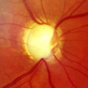
Glaucomatous optic nerve
This information does not replace an individual medical assessment. Follow the advice of the team looking after you.
Open article on its own page Which symptoms should I watch for if glaucoma is suspected?
Unfortunately, none.
The vast majority of glaucomas are completely symptom-free. Several factors contribute to this:
1) Ocular hypertension, a major risk factor for the development and progression of glaucoma, usually causes no symptoms.
2) Visual function defects are initially peripheral and usually do not affect the detailed central vision we tend to notice most.
3) Several compensatory sensory mechanisms mask our perception of visual loss.
Watch video on YouTube ↗Original video in PortugueseThis information does not replace an individual medical assessment. Follow the advice of the team looking after you.
Open article on its own page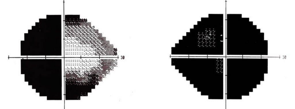 I have relatives who are blind from glaucoma. Is the same likely to happen to me?
Not necessarily.
Many factors help determine the prognosis of glaucoma. Because it is a symptom-free disease in which established damage is irreversible, early diagnosis is essential to preserve lifelong visual function. Heredity is a risk factor for developing the disease, but descendants are usually diagnosed earlier, which is highly beneficial for preserving their sight.
This information does not replace an individual medical assessment. Follow the advice of the team looking after you.
Open article on its own pageIs open-angle or angle-closure glaucoma worse?
There is no universal rule. Around 75% of glaucoma is open-angle, with the rest being narrow-angle or angle-closure. Although less common, narrow-angle glaucoma can be more destructive and accounts for around 50% of blindness cases. Secondary angle-closure glaucoma has a worse prognosis, but progression in many cases of primary narrow-angle glaucoma can be prevented by surgically improving the anatomy of the angle.
This information does not replace an individual medical assessment. Follow the advice of the team looking after you.
Open article on its own pageCan laser treatment or surgery cure my glaucoma?
Generally, no. Laser and surgical treatments aim to optimise control of intraocular pressure, thereby limiting glaucoma progression so that it does not threaten the patient's visual function. These techniques cannot, however, restore function already lost to glaucoma.
In certain cases of anatomical narrow-angle glaucoma, lens surgery can definitively resolve the problem.
This information does not replace an individual medical assessment. Follow the advice of the team looking after you.
Open article on its own pageCan vision or visual field lost to glaucoma be restored?
Vision loss caused by optic nerve damage in glaucoma is irreversible. Available treatments aim to control intraocular pressure and reduce the risk of worsening; they do not restore vision already lost.
Early diagnosis, regular follow-up and adherence to treatment are essential to preserve vision.
Further information — National Eye Institute ↗
This information does not replace an individual medical assessment. Follow the advice of the team looking after you.
Open article on its own page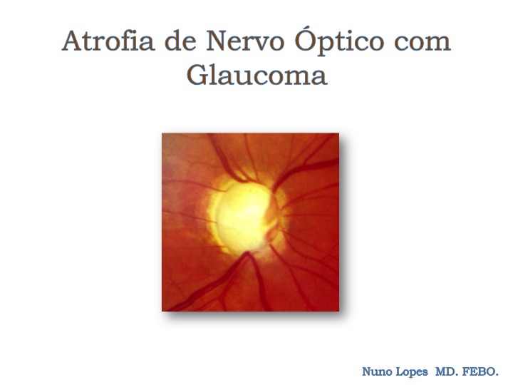 Does having glaucoma mean I will go blind?
No.
Glaucoma is undoubtedly a major problem nationally and worldwide, but fortunately blindness affects only a minority of treated patients.
Successful treatment rests on three main foundations:
1) Early diagnosis
2) Controlling progression through optimised treatment and appropriate follow-up
3) Following treatment as prescribed (treatment adherence)
This information does not replace an individual medical assessment. Follow the advice of the team looking after you.
Open article on its own page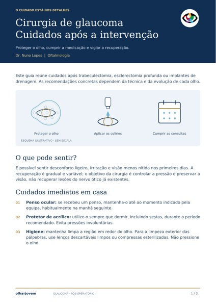 Care after glaucoma surgery
After glaucoma surgery, protecting the eye, applying medication correctly and attending review appointments are all part of recovery.
Specific care depends on the technique used and your recovery. Always confirm which drops to use in the operated eye, as the schedule may change after the procedure.
Care and follow-up
- Do not rub or press on the eye. Wear the shield while sleeping for the advised period.
- Follow your team's restrictions on water, exertion, driving and work.
- Bring all your eye drops and medicines to follow-up appointments.
When to seek help
Severe pain, sudden or significant reduction in vision, increasing redness or pus-like discharge require immediate contact with the team or urgent assessment.
PATIENT GUIDE
After glaucoma surgery
Protection, medication and follow-up after trabeculectomy, deep sclerectomy or drainage implants.
This information does not replace medical assessment. Follow your team's individual instructions.
Open article on its own pageCataracts and intraocular lenses
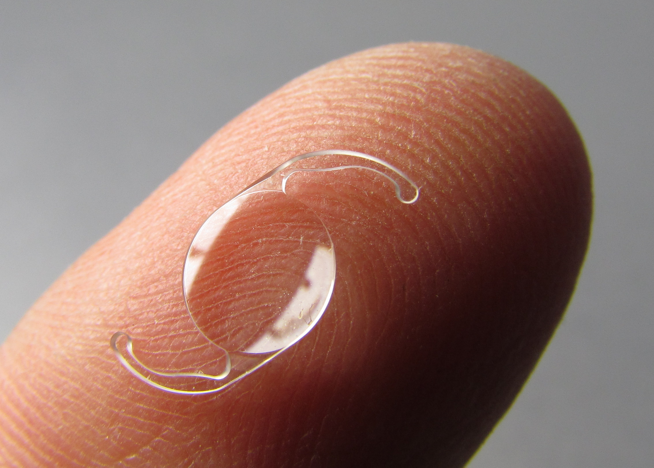 Which lens is best for my cataract or lens surgery?
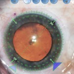
Lens surgery offers an opportunity to correct the eye's pre-existing prescription. Lenses of various powers are available, with spherical, aspheric, toric, multifocal and other characteristics…
It is therefore now possible to correct refractive errors such as myopia, hyperopia, astigmatism and presbyopia.
The ideal choice depends on the patient's and clinician's decision after consultation and careful assessment of additional tests.
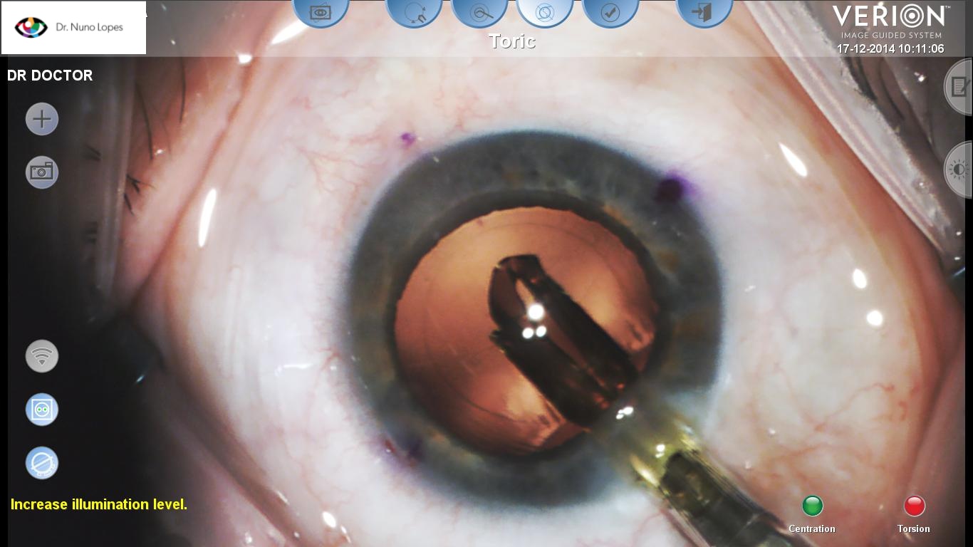
Intraocular lens implantation
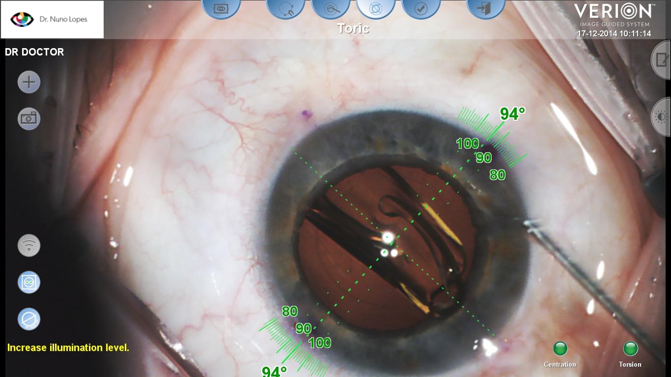
Intraocular lens implantation
This information does not replace an individual medical assessment. Follow the advice of the team looking after you.
Open article on its own pageAstigmatism and lens choice in lens surgery
Correcting astigmatism with toric lenses and comparing monofocal, extended depth of focus and trifocal lenses.
Read article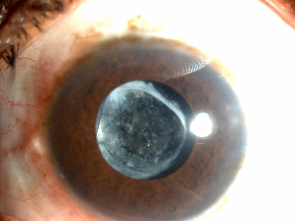 What does it mean to have a “dirty” intraocular lens?
The expression “a dirty intraocular lens” is sometimes used to describe clouding of the back of the capsular bag in which the lens was implanted during lens surgery.
To understand this condition, it helps first to clarify a few anatomical concepts.
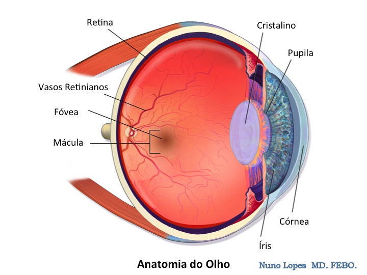
The crystalline lens is the natural lens inside the eye that helps us focus on objects. It lies centrally, separating the front and back parts of the eye.
Anatomically, the natural lens consists of flexible, protein-rich material enclosed in a capsule. Equatorial fibres (zonules) connect this capsule to the ciliary muscle, whose action allows young people to change the lens's curvature and focus sharply on near objects.
When lens surgery is indicated for cataract, a high refractive error and/or presbyopia, the inner contents are broken up and aspirated, while the surrounding capsular bag should remain intact.
After an intraocular lens has been implanted, the capsular bag can therefore still undergo fibrosis and clouding that may significantly reduce visual quality.
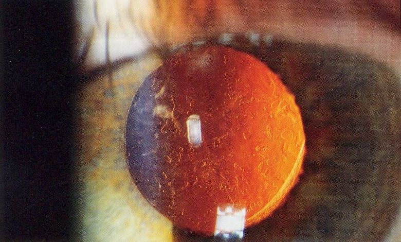
Capsular opacification
Posterior capsule opacification is treated by a capsulotomy in a single outpatient YAG (Yttrium Aluminum Garnet) laser session, with rapid visual recovery.
This information does not replace an individual medical assessment. Follow the advice of the team looking after you.
Open article on its own page After cataract surgery
, follow the individual instructions given by your surgical team.
Care of the operated eye
- Wash your hands before applying eye drops.
- Do not rub or press on the operated eye.
- Keep water out of the eye and wait for approval before swimming, physical exertion or driving again.
- Use eye protection as instructed and attend your follow-up appointment.
Medication
Use eye drops exactly as prescribed. The medicine, frequency and duration of treatment are determined individually.
When to seek help
Contact the medical team promptly if you have increasing pain, worsening redness, reduced vision or discharge. Flashes, new shadows or floaters also need assessment.
TO CONSULT AT HOME
Instructions after cataract surgery
Read Dr. Nuno Lopes's leaflet about care after surgery. Medication must always follow the individual prescription you received.
PATIENT GUIDE
Complete cataract guide
Learn about cataracts, preparing for surgery and care during recovery.
This information does not replace an individual medical assessment. Follow the advice of the team looking after you.
Open article on its own pageEveryday care
 Vision and sport: see better, play better
Visual assessment, correction and eye protection tailored to the demands of each sport.
Read article How do I apply eye drops or ointment?
• Wash your hands
• Sit or lie down with your head tilted back
• Gently lower the bottom eyelid and apply the drop or ointment
Close the eye for 2 to 3 minutes and wipe away any excess with a clean tissue
This information does not replace an individual medical assessment. Follow the advice of the team looking after you.
Open article on its own page Information for contact lens wearers
Read the care guide for contact lens wearers.
CARE GUIDE
Contact lenses
View the document or save a copy to read later.
This information does not replace an individual medical assessment. Follow the advice of the team looking after you.
Open article on its own page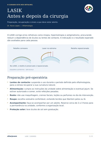 Care before and after LASIK
Read the LASIK preparation and recovery guide. These instructions are specific to this technique; PRK requires its own care.
PATIENT GUIDE
LASIK care guide
Preparation, recovery and warning signs. This guide is specific to LASIK; PRK instructions are provided individually.
Vision, driving and support
 Driving licence
Vision assessment forms part of the assessment of fitness to drive. When necessary, an ophthalmology consultation can document visual function and clarify limitations relevant to driving.
The form below is intended to record the eye assessment and must be completed by the doctor during the consultation.
REFERENCE DOCUMENT
Ophthalmology report
Driving licence renewal form, with fields for the visual assessment, location, date, signature and medical identification label.
This information does not replace an individual medical assessment. Follow the advice of the team looking after you.
Open article on its own page Low vision – Disability
Information on visual disability and resources to support independence, rehabilitation and inclusion for blind and partially sighted people.
REFERENCE DOCUMENTS
Portuguese National Disability Assessment Table
View the full document in your browser or save a copy to read later.
Support networks in Portugal
These organisations provide information and support for people with visual disabilities and their families. Contact them to find out which services are appropriate for your situation.
ACAPO
Associação dos Cegos e Amblíopes de Portugal. Support for independence, habilitation and rehabilitation, inclusion and protection of the rights of people with visual disabilities.
AAICA
Associação de Apoio e Informação a Cegos e Amblíopes. Accessible information and guidance for blind and partially sighted people and their families.
About AAICA ↗Centro de Reabilitação Nossa Senhora dos Anjos
A Santa Casa da Misericórdia de Lisboa service for people with acquired blindness or low vision, supporting independence, orientation and mobility, and activities of daily living.
Explore rehabilitation programmes ↗Rights and public support
Official guide with information on rights, social support, employment, training and access to the Balcões da Inclusão support network.
Read the guide on gov.pt ↗This information does not replace an individual medical assessment. Follow the advice of the team looking after you.
Open article on its own pageNo articles found for this search. Try another term or choose another topic.
Your question deserves attention.
For advice about your situation, book an ophthalmology appointment.
View consultation locations ↗