


Allergic conjunctivitis
Itching is the hallmark symptom of eye allergy.
Read article ↗
Eye conditions · 3 MIN READ
WHAT YOU NEED TO KNOW
The cornea is the clear window at the front of the eye and does much of its focusing. In keratoconus, the cornea becomes thinner and weaker and gradually bulges into a cone. The image becomes distorted and an irregular astigmatism develops, which glasses correct less and less well.
THE CORNEA IN PROFILE
Why the image becomes distorted
A regular cornea focuses light to a point; a cone-shaped cornea scatters it.
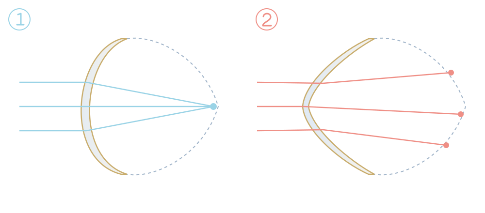
Regular curvature: light converges to a point and the image is sharp.
The cornea thins and bulges into a cone: light scatters and the image becomes blurred and distorted.
CLINICAL PHOTOS
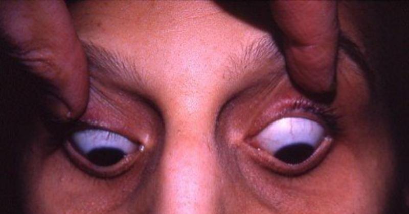
Advanced keratoconus: when looking down, the cone-shaped cornea bulges the edge of the lower eyelid (Munson’s sign).
Photo: William Charles Caccamise, Sr, MD · CC BY-SA 4.0 · Wikimedia Commons
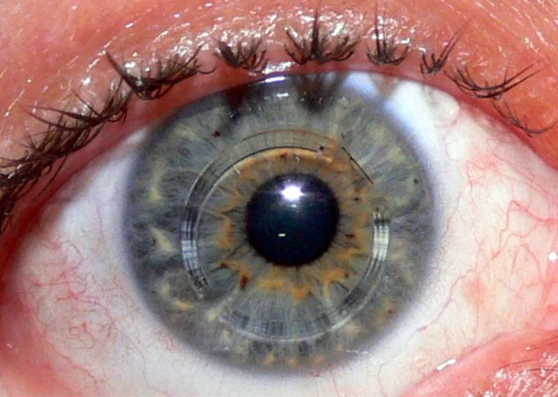
Intracorneal ring segments nine days after surgery in an eye with keratoconus: the two clear arcs sit within the thickness of the cornea, around the pupil, and help to regularise its shape.
Photo: Edouard Spooner · CC BY-SA 2.0 · Wikimedia Commons
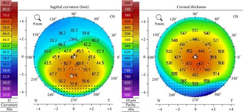
Topography of a keratoconus (stage II): on the left, the curvature map shows the steepest area (orange and red) below the centre; on the right, the thickness map shows the thinnest cornea at the same spot.
Photo: Elise A. Slim et al. · CC BY 4.0 · Wikimedia Commons
Rarely, in advanced cases, the cornea can suddenly fill with fluid, with rapid loss of vision, pain and a white patch on the cornea. This should be examined urgently.
In the early stages, keratoconus may not be visible on a routine examination. Corneal topography and tomography map the curvature and thickness at every point, allow early diagnosis and show whether it is progressing by comparing scans over time. That is why these tests are also done before any laser refractive surgery.
Treatment has two aims: to halt progression and to correct vision.
People with keratoconus should not have LASIK, because it weakens the cornea further. Regular follow-up with corneal tomography is essential, especially in younger patients.
Learn more:
Refractive errors: myopia, hyperopia and astigmatism
This information does not replace an individual medical assessment. Follow the advice of the team looking after you.
Clinical responsibility: Dr. Nuno Lopes · Ophthalmology
Page update:
This date refers to the page presentation and organisation.
Clinical review: Dr. Nuno Lopes ·
CONTINUE READING
Itching is the hallmark symptom of eye allergy.
Read article ↗
A refractive error exists when light entering the eye is not focused on the retina.
Read article ↗
The laser reshapes the cornea to correct short-sightedness, long-sightedness and astigmatism and reduce dependence on glasses or contact lenses.
Read article ↗