


Diabetic retinopathy
Diabetic retinopathy is the most common eye complication of diabetes.
Read article ↗
Eye conditions · 2 MIN READ
WHAT YOU NEED TO KNOW
Retinal veins carry blood out of the eye. When one becomes blocked, blood cannot drain: bleeding and fluid appear in the retina. If the main vein is affected (central occlusion), the whole retina suffers; if a branch is affected, only part of it.
THE BACK OF THE EYE
What the ophthalmologist sees
In a branch occlusion, the bleeding follows the area drained by the blocked vein.
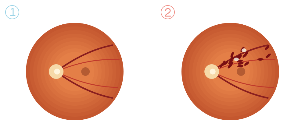
The veins, darker and wider, drain blood towards the optic nerve.
Blood is trapped: haemorrhages, white spots and fluid appear in the retina.
Blurred vision or a patch in your field of vision, in one eye, appearing suddenly or on waking, without pain. A small branch occlusion may go unnoticed.
It is more common after the age of 60 and is linked to cardiovascular risk factors: high blood pressure, diabetes, high cholesterol and smoking. Glaucoma and raised eye pressure also increase the risk. So, as well as treating the eye, a check-up with your family doctor or an internal medicine specialist is important.
This information does not replace an individual medical assessment. Follow the advice of the team looking after you.
Clinical responsibility: Dr. Nuno Lopes · Ophthalmology
Page update:
This date refers to the page presentation and organisation.
CONTINUE READING
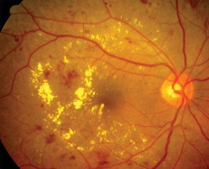
Diabetic retinopathy is the most common eye complication of diabetes.
Read article ↗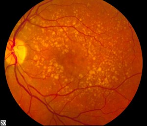
AMD affects the macula, the part of the retina responsible for central, detailed vision.
Read article ↗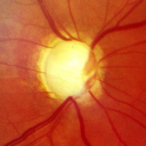
Glaucoma damages the optic nerve and, without treatment, causes vision loss that cannot be recovered.
Read article ↗