


Retinal vein occlusion
A retinal vein is blocked, blood builds up and the retina swells.
Read article ↗
Eye conditions · 2 MIN READ
WHAT YOU NEED TO KNOW
The optic nerve carries visual information from the eye to the brain. In ischaemic optic neuropathy, the blood supply to the part of the nerve inside the eye drops suddenly and the nerve fibres are damaged. The result is sudden, painless vision loss in one eye, often noticed on waking, frequently in the upper or lower half of the field of vision.
THE OPTIC NERVE AND THE VISUAL FIELD
A swollen nerve and half the field lost
In the acute phase, the optic disc looks swollen; vision loss often affects the upper or lower half.
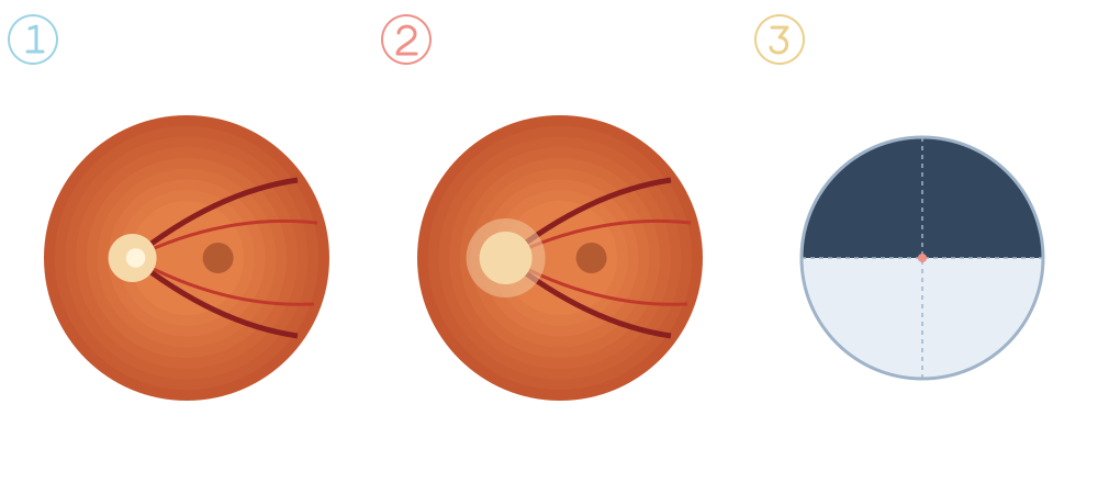
Sharp edges, where the nerve fibres leave the eye.
The lack of blood flow makes the disc swell; over time it becomes pale.
Typical loss of the upper or lower half of vision in that eye.
The most common form (non-arteritic) is associated with high blood pressure, diabetes, cholesterol, sleep apnoea and a small optic nerve. The rarer arteritic form is caused by giant cell arteritis, an inflammation of the arteries occurring after 50. This form can affect the other eye within days and needs immediate treatment with corticosteroids.
In the non-arteritic form, no treatment has been proven to restore lost vision, although some patients improve somewhat over the following months. The aim is to protect the other eye: control blood pressure, diabetes and cholesterol, treat sleep apnoea, stop smoking and review with your doctor any medicines that lower blood pressure a lot at night.
This information does not replace an individual medical assessment. Follow the advice of the team looking after you.
Clinical responsibility: Dr. Nuno Lopes · Ophthalmology
Page update:
This date refers to the page presentation and organisation.
Clinical review: Dr. Nuno Lopes ·
CONTINUE READING

A retinal vein is blocked, blood builds up and the retina swells.
Read article ↗
Glaucoma damages the optic nerve and, without treatment, causes vision loss that cannot be recovered.
Read article ↗
Diabetic retinopathy is the most common eye complication of diabetes.
Read article ↗